is the headline sign.
Symptoms, risk, screening and diagnostic confirmation
Detect Bladder Cancer is about choosing the right test for the right risk.
A bladder cancer detection guide covering visible blood in urine, cystoscopy, urine cytology, blue-light cystoscopy, CT urogram and biopsy.
directly sees the bladder.
helps for high-grade disease.
evaluates upper tracts.
Brief outline
What bladder cancer means.
Bladder cancer usually presents with hematuria. Cystoscopy and biopsy are central because urine tests cannot safely rule it out.
Detection frame
Screening is for people without symptoms. Diagnosis is for symptoms, abnormal examination, family history, imaging findings or abnormal labs. The same test can be sensible in one setting and misleading in another.
Do not ignore
Symptoms and signs not to ignore.
Risk and genetics
Demographics, risks and genetics.
Screening
Who should actually be screened?
No broad average-risk screening program exists. High-risk occupational or prior cancer surveillance is different.
Cystoscopy
Direct inspection of bladder mucosa.
TURBT/biopsy
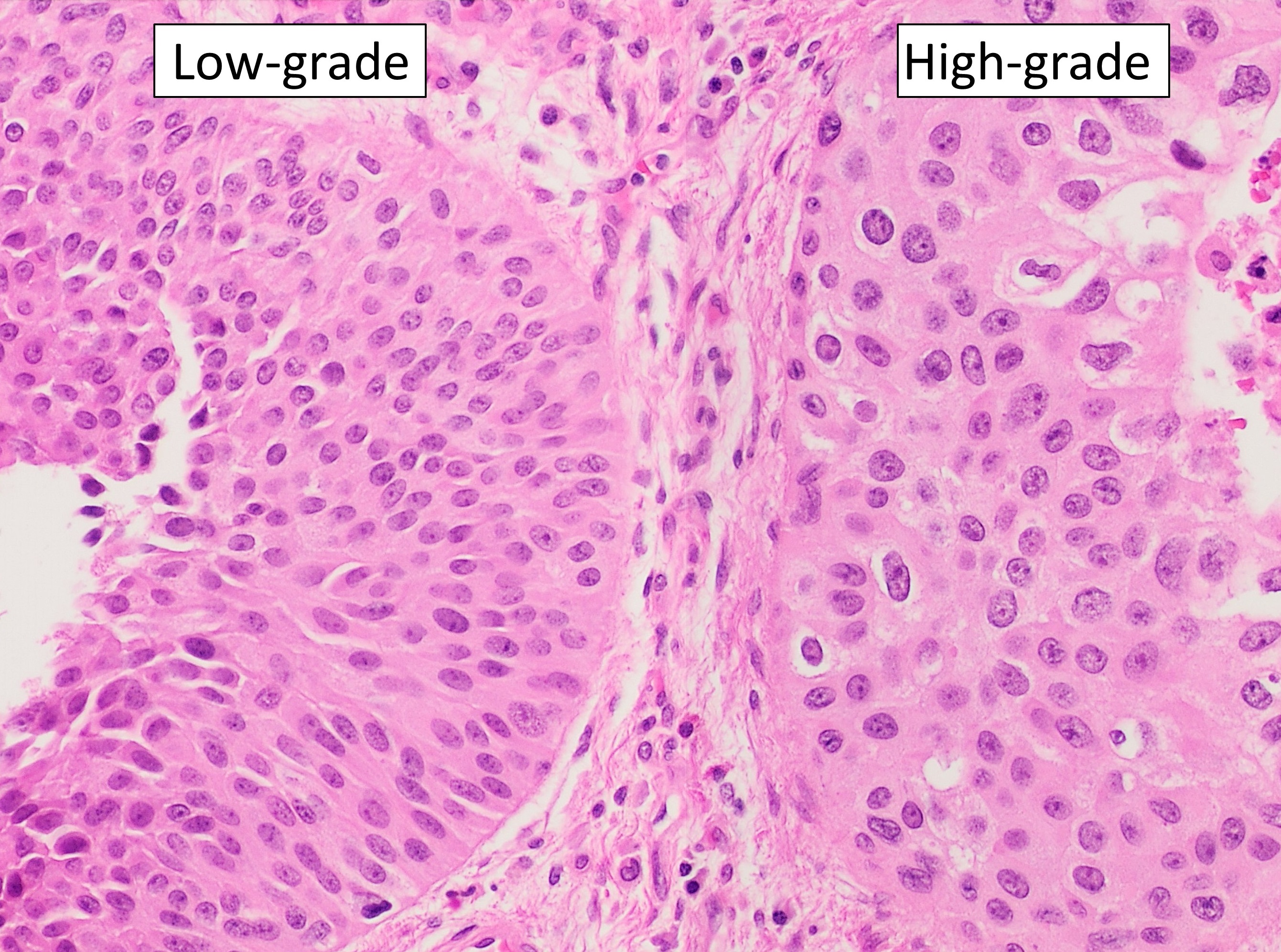
Confirms grade, invasion and muscle involvement.
Cytology
Good for high-grade/CIS, poor for many low-grade tumors.
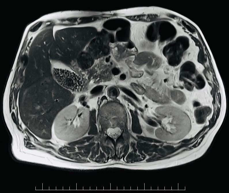
CT urogram
Evaluates kidneys, ureters and bladder.
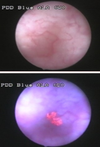
Blue-light cystoscopy
Can improve CIS/tumor detection in selected settings.
Urine markers
Adjuncts only; false positives and negatives occur.
Images, labs and tissue
Useful tests and what they can miss.
Detection usually needs a chain: suspicion, imaging or lab triage, tissue or cytology when needed, then pathology and molecular testing where it changes management.
Pros, cons and controversy
Benefits, harms and the false-positive problem.
The fatal mistake is assuming hematuria is infection, prostate or anticoagulants without follow-up. The counter-risk is expensive urine marker panels that still require cystoscopy.
Sources and trials