with contrast is the key diagnostic imaging test.
Seizures, MRI, CT and molecular pathology
Brain tumors are usually detected because symptoms force imaging, not because screening finds them.
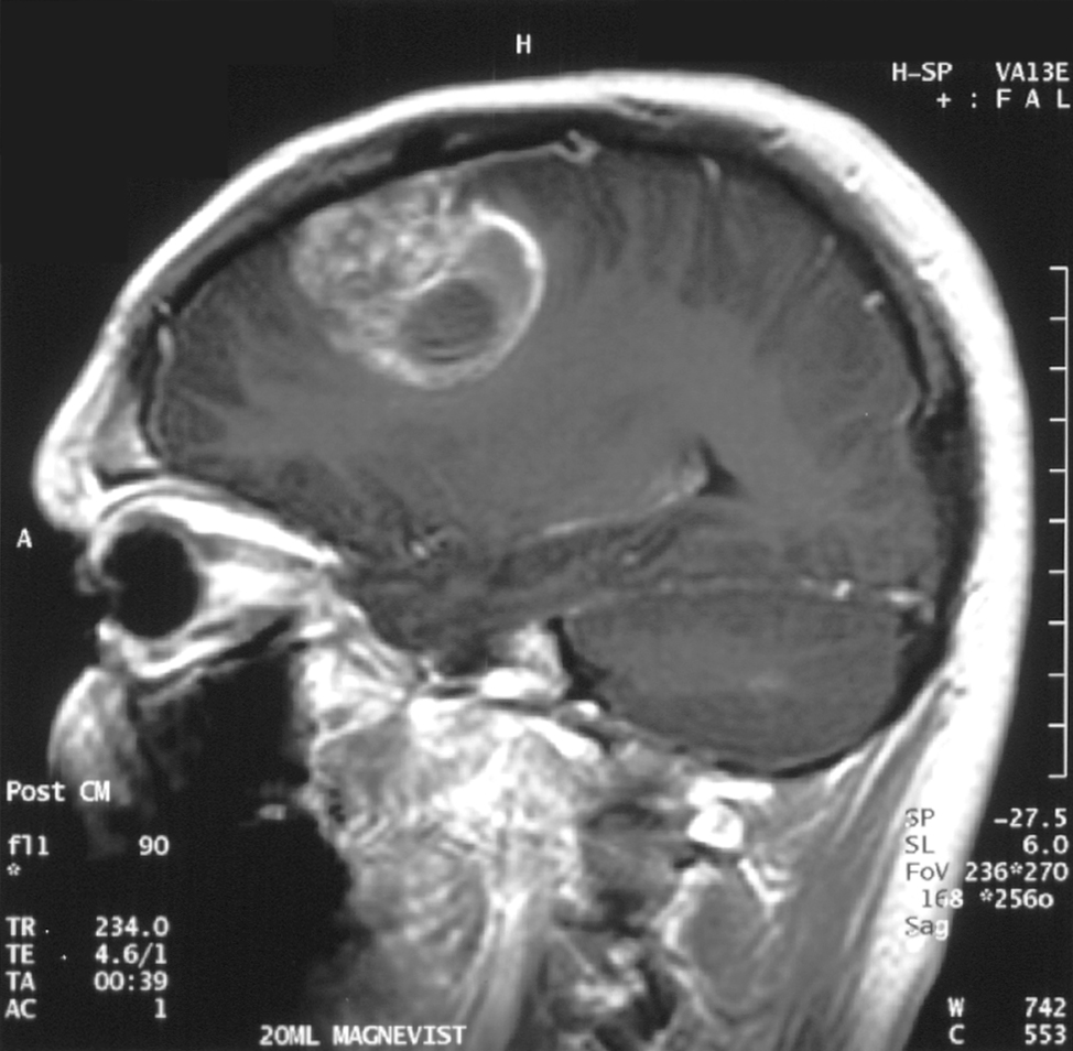
There is no routine average-risk brain cancer screening. New seizure, progressive neurologic deficit or worrying headache pattern should lead to urgent imaging, usually MRI with contrast.
is often first in emergency settings.
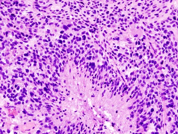
or resection provides histology and molecular markers.
routine population screening is recommended.
Brief outline
Primary brain tumors differ from brain metastases.
Gliomas include astrocytoma, oligodendroglioma and glioblastoma, now classified by molecular markers such as IDH and 1p/19q. Metastases from lung, breast, melanoma, kidney and other cancers are more common than many primary tumors.
Detection frame
Detection starts with recognizing neurologic red flags and ordering the right imaging. Tissue diagnosis increasingly means molecular diagnosis, not just microscope appearance.
Do not ignore
New neurologic symptoms are the screening test you actually have.
Risk and genetics
Most brain tumors have no obvious preventable cause.
Screening
Average-risk screening is not recommended.
Whole-body MRI usually does not include a dedicated brain MRI good enough for every subtle lesion, and even brain MRI screening would create incidental findings. High-risk syndromes need syndrome-specific surveillance.
CT brain
Fast first test for acute neurologic symptoms, hemorrhage or mass effect.
MRI with contrast
Best standard test for suspected brain tumor.
Functional MRI/tractography
Selected cases near speech or motor pathways.
Biopsy or resection
Confirms tumor type and molecular profile.
CSF testing
Useful for lymphoma, leptomeningeal disease or infection mimics, not routine for every mass.
Liquid biopsy/CSF DNA
Promising but not a population screening replacement.
Images, labs and tissue
MRI pattern guides urgency, but tissue/molecular diagnosis guides treatment.
CT is fast for bleeding, mass effect and emergency triage. MRI with contrast better defines tumor, edema, infiltration and surgical planning. Biopsy/resection provides histology, IDH, MGMT and other molecular markers.
Pros, cons and controversy
The controversy is incidentalomas and false reassurance.
A scan can find meningiomas, aneurysms, white-matter changes or benign cysts that create anxiety. A normal non-dedicated scan can also falsely reassure someone with evolving symptoms. Symptom-led MRI remains the practical pathway.
Sources and trials