symptoms overlap with gallstones.
Symptoms, risk, screening and diagnostic confirmation
Detect Gallbladder Cancer is about choosing the right test for the right risk.
A gallbladder cancer detection guide covering gallstones, porcelain gallbladder, jaundice, ultrasound, CT/MRI, MRCP and incidental pathology after cholecystectomy.
is usually first.
stage suspicious masses.
may find incidental cancer after surgery.
Brief outline
What gallbladder cancer means.
Gallbladder cancer is uncommon and often found late or incidentally after gallbladder removal for presumed benign disease.
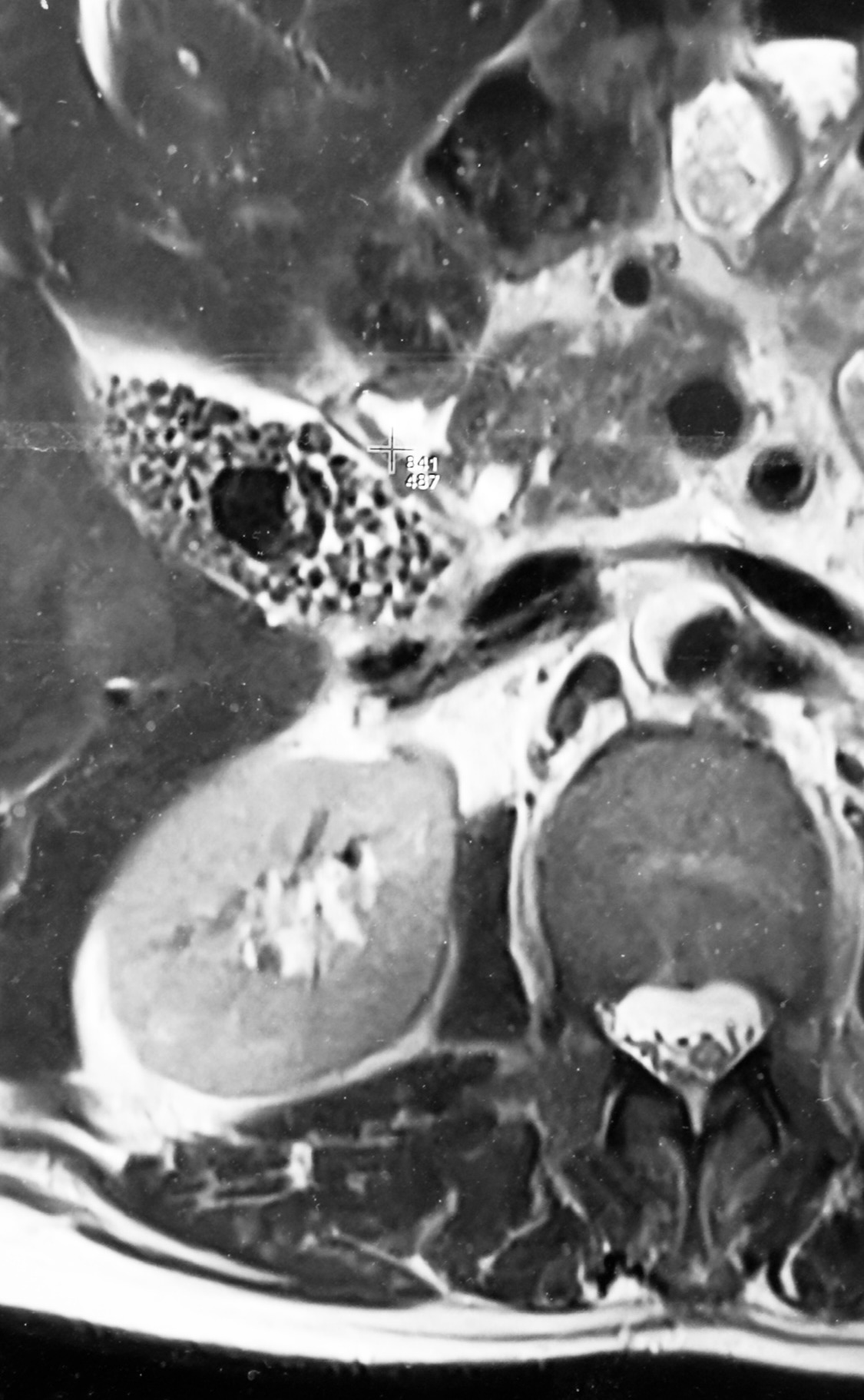
Detection frame
Screening is for people without symptoms. Diagnosis is for symptoms, abnormal examination, family history, imaging findings or abnormal labs. The same test can be sensible in one setting and misleading in another.
Do not ignore
Symptoms and signs not to ignore.
Risk and genetics
Demographics, risks and genetics.
Screening
Who should actually be screened?
There is no average-risk screening. Surveillance or surgery decisions for gallbladder polyps depend on size, growth and risk factors.
Ultrasound
Finds stones, wall thickening, polyps or mass.
CT abdomen/chest
Assesses liver invasion, nodes and spread.
MRI/MRCP
Defines biliary obstruction and local anatomy.
EUS/ERCP brushings
Selected cases with bile duct involvement.
Surgical pathology
Often the diagnostic moment for incidental cancer.
Gallstone assumption
Gallstones can coexist with cancer.
Images, labs and tissue
Useful tests and what they can miss.
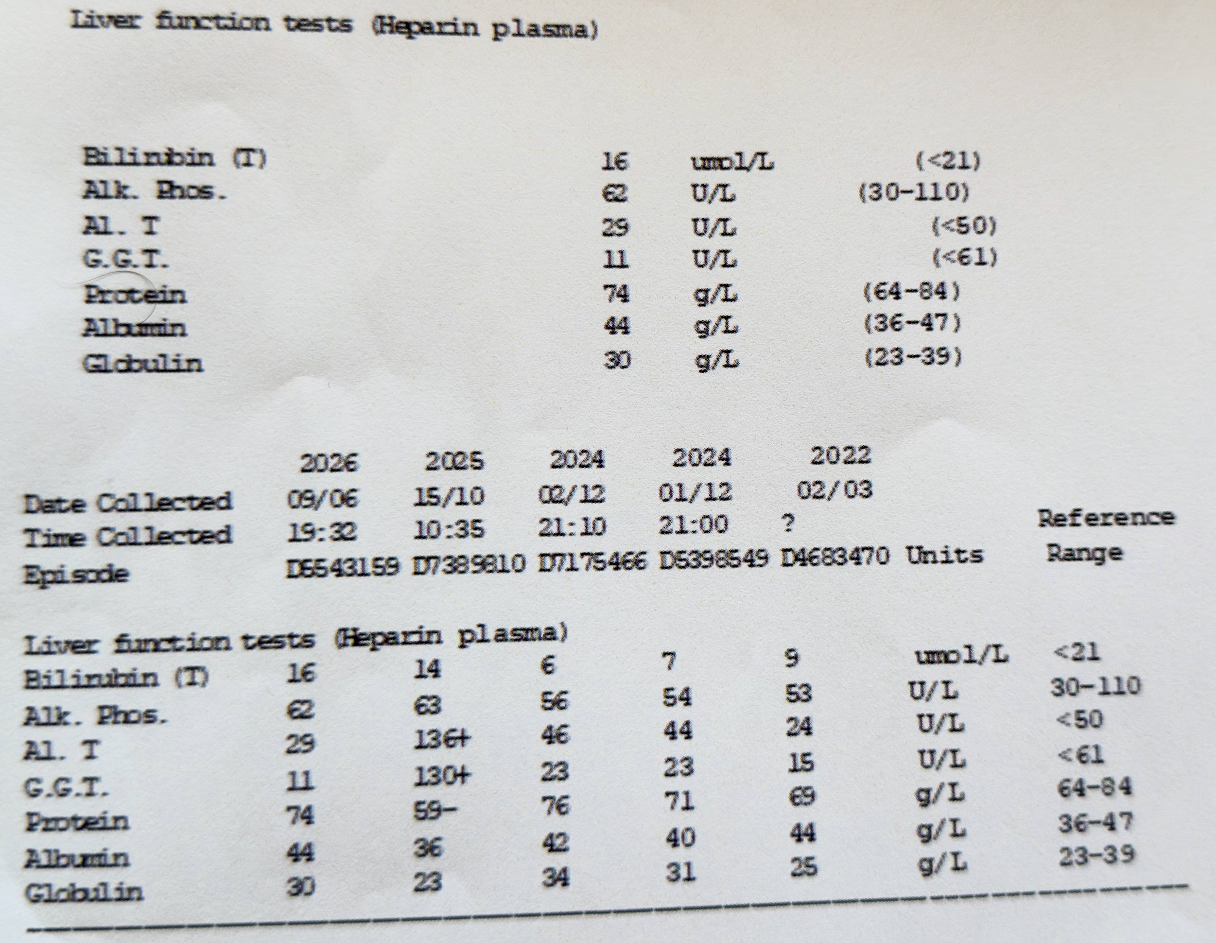
Detection usually needs a chain: suspicion, imaging or lab triage, tissue or cytology when needed, then pathology and molecular testing where it changes management.
Pros, cons and controversy
Benefits, harms and the false-positive problem.
Most gallbladder pain is benign gallstone disease, but painless jaundice or a mass is different. Finding frail patients with advanced disease also raises hard questions about benefit.
Sources and trials