often reveal functioning adenomas.
Symptoms, risk, screening and diagnostic confirmation
Detect Pituitary Adenoma is about choosing the right test for the right risk.
A pituitary adenoma detection guide covering prolactin, cortisol, acromegaly, visual field loss, pituitary MRI and incidentaloma follow-up.
pituitary protocol is key.
detect chiasm compression.
sudden headache/vision loss is emergency.
Brief outline
What pituitary adenoma means.
Pituitary adenomas are usually benign tumors, but they can seriously harm through hormone excess/deficiency, optic chiasm compression or pituitary apoplexy.
Detection frame
Screening is for people without symptoms. Diagnosis is for symptoms, abnormal examination, family history, imaging findings or abnormal labs. The same test can be sensible in one setting and misleading in another.
Do not ignore
Symptoms and signs not to ignore.
Risk and genetics
Demographics, risks and genetics.
Screening
Who should actually be screened?
No population screening exists. Hormone syndromes, visual symptoms and incidental MRI findings drive detection.
Prolactin, IGF-1, cortisol/ACTH axis
Chosen by clinical pattern and incidentaloma workup.
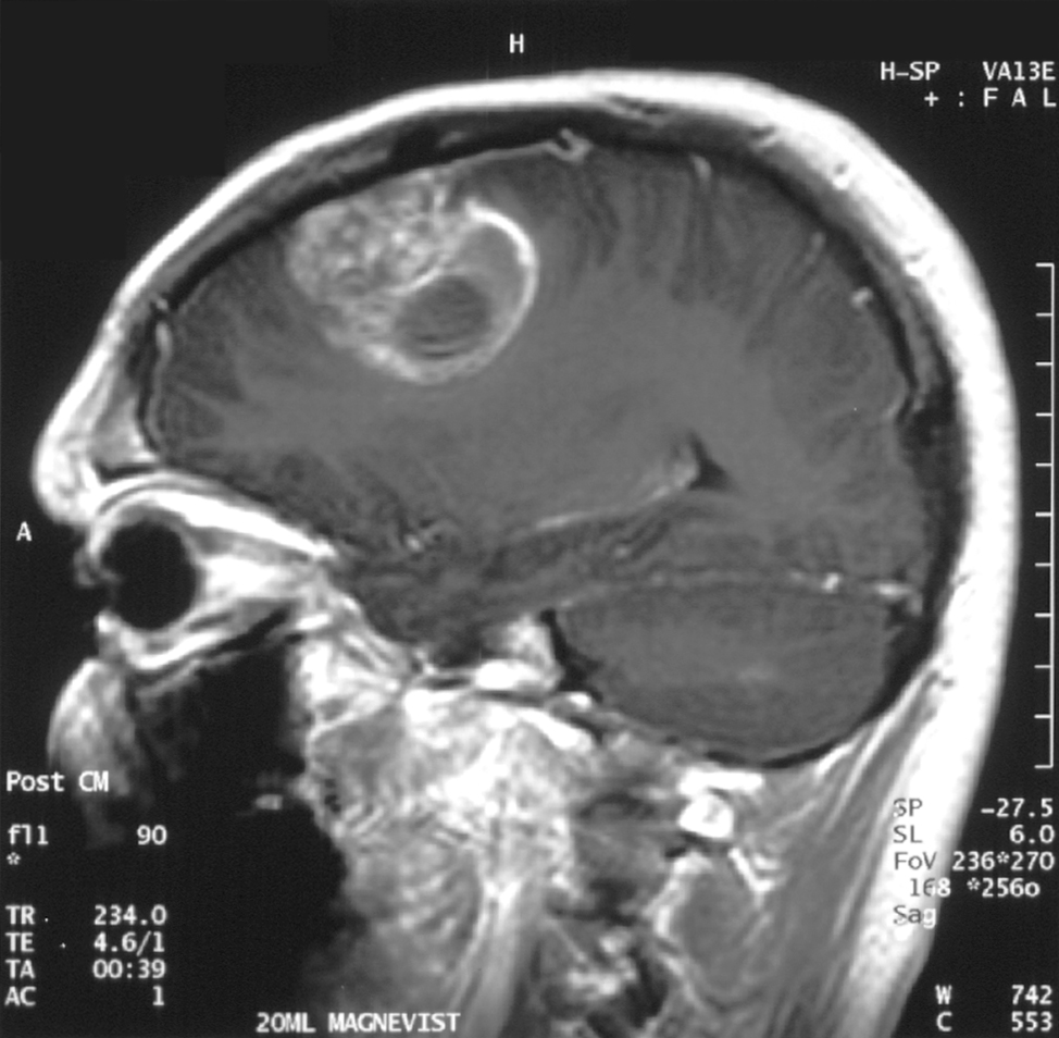
Pituitary MRI with contrast
Defines microadenoma/macroadenoma and chiasm compression.
Formal visual fields
Essential if near optic chiasm.
Pituitary function panel
Checks adrenal, thyroid and gonadal axes.
Urgent MRI/CT and steroids
For suspected apoplexy with adrenal risk.
Tiny incidentalomas
Often need structured follow-up, not panic.
Images, labs and tissue
Useful tests and what they can miss.
Detection usually needs a chain: suspicion, imaging or lab triage, tissue or cytology when needed, then pathology and molecular testing where it changes management.

Pros, cons and controversy
Benefits, harms and the false-positive problem.
These are 'benign' but not harmless. The false-positive problem is MRI incidentalomas; the real danger is missing hormone excess or optic compression.
Sources and trials