but important in children.
Symptoms, risk, screening and diagnostic confirmation
Detect Rhabdomyosarcoma is about choosing the right test for the right risk.
A rhabdomyosarcoma detection guide covering childhood soft-tissue masses, head and neck symptoms, urinary/genital symptoms, MRI, CT, biopsy and FOXO1 fusion testing.
maps local extent.
confirms subtype.
fusion status affects risk.
Brief outline
What rhabdomyosarcoma means.
Rhabdomyosarcoma is a rare soft-tissue sarcoma, usually in children, arising from skeletal-muscle lineage cells but able to appear in many body sites.
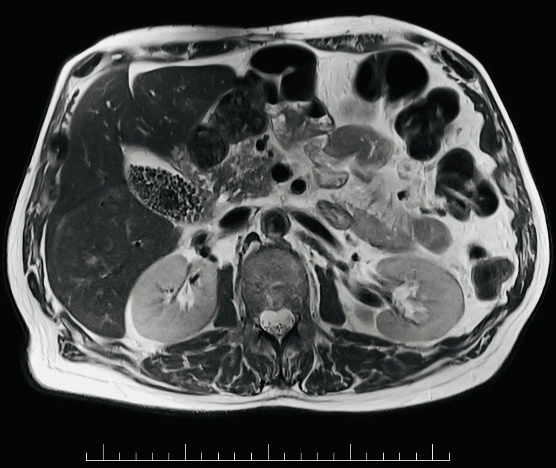
Detection frame
Screening is for people without symptoms. Diagnosis is for symptoms, abnormal examination, family history, imaging findings or abnormal labs. The same test can be sensible in one setting and misleading in another.
Do not ignore
Symptoms and signs not to ignore.
Risk and genetics
Demographics, risks and genetics.
Screening
Who should actually be screened?
No screening program exists. Detection is symptom-led, with low threshold for imaging persistent growing masses in children.
MRI
Best maps local soft-tissue extent.
CT chest/PET/bone marrow selected
Assesses spread depending on risk.
Core/open biopsy
Planned by sarcoma team.
IHC and fusion testing
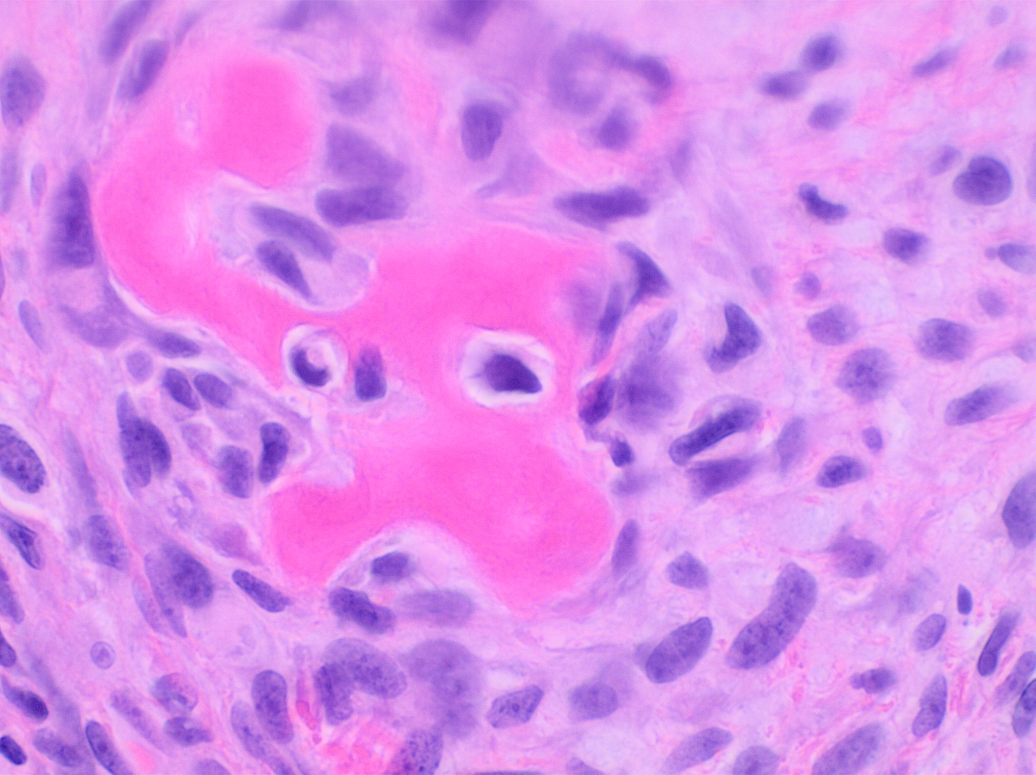
Confirms RMS subtype and risk.
Syndrome assessment
Consider with family history or syndromic features.
Treating as infection only
Persistent masses after antibiotics need imaging.
Images, labs and tissue
Useful tests and what they can miss.
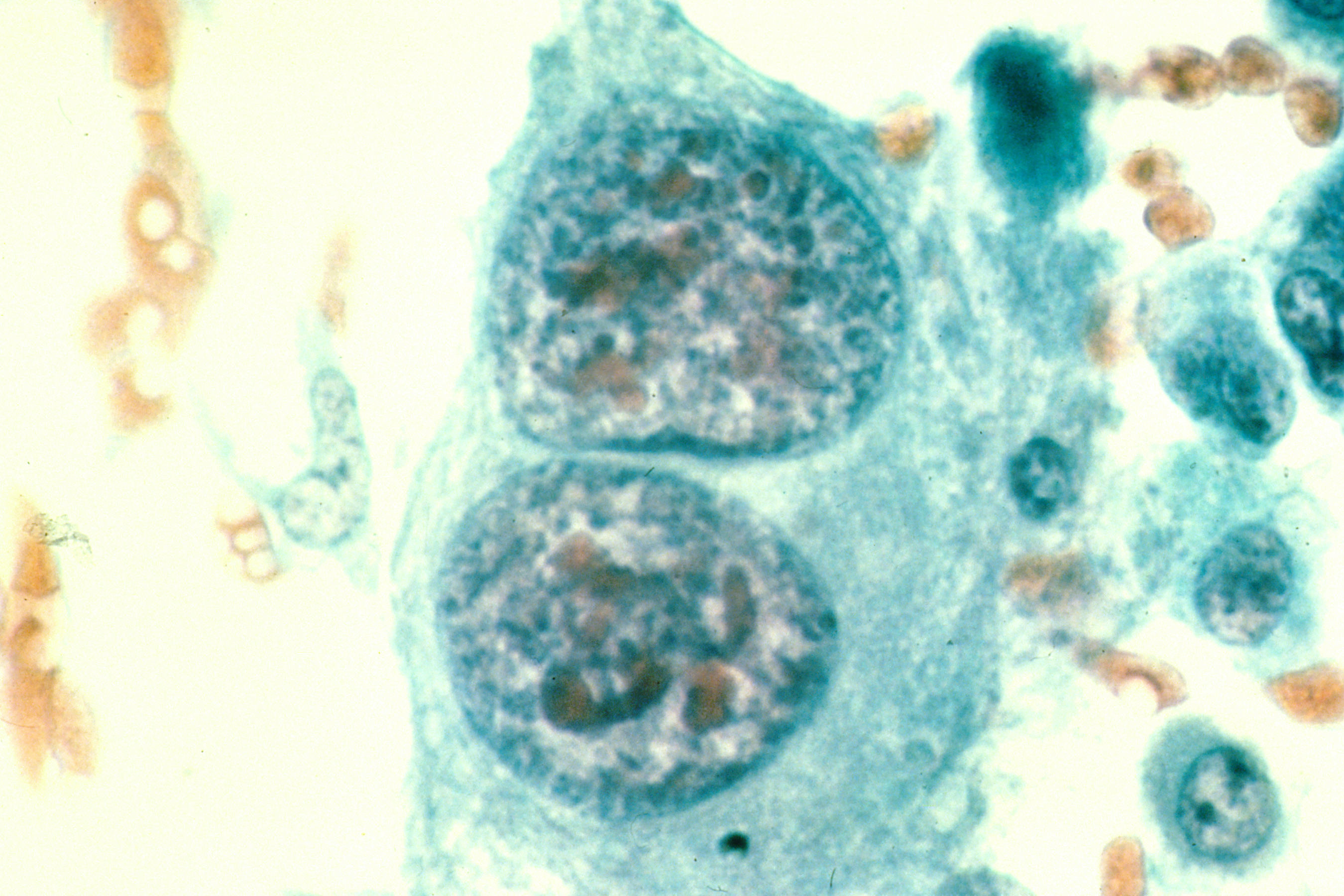
Detection usually needs a chain: suspicion, imaging or lab triage, tissue or cytology when needed, then pathology and molecular testing where it changes management.
Pros, cons and controversy
Benefits, harms and the false-positive problem.
Because it is rare, delay is easy. The main safety message is that persistent, growing or unusual pediatric masses deserve imaging and planned biopsy.
Sources and trials