commonest solid cancer in younger adult men.
Symptoms, risk, screening and diagnostic confirmation
Detect Testicular Cancer is about choosing the right test for the right risk.
A testicular cancer detection guide covering painless lump, ultrasound, AFP, beta-hCG, LDH, CT staging and orchiectomy histology.
key first imaging test.
AFP, hCG and LDH help subtype/stage.
radical inguinal orchiectomy is diagnostic/therapeutic.
Brief outline
What testicular cancer means.
Testicular cancer is often curable, especially when detected early. A solid testicular mass is cancer until proven otherwise.
Detection frame
Screening is for people without symptoms. Diagnosis is for symptoms, abnormal examination, family history, imaging findings or abnormal labs. The same test can be sensible in one setting and misleading in another.
Do not ignore
Symptoms and signs not to ignore.
Risk and genetics
Demographics, risks and genetics.
Screening
Who should actually be screened?
No formal population screening program exists, but self-awareness and prompt ultrasound for a lump are sensible.
Scrotal ultrasound
Distinguishes intratesticular mass from cyst/epididymal disease.
AFP, beta-hCG, LDH
Markers help diagnosis, staging and follow-up.
CT abdomen/pelvis/chest
Assesses nodal and metastatic disease.
Radical inguinal orchiectomy
Provides diagnosis and local treatment.
Seminoma vs non-seminoma
Changes treatment and marker interpretation.
Trans-scrotal biopsy
Generally avoided because it can alter spread pathways.
Images, labs and tissue
Useful tests and what they can miss.
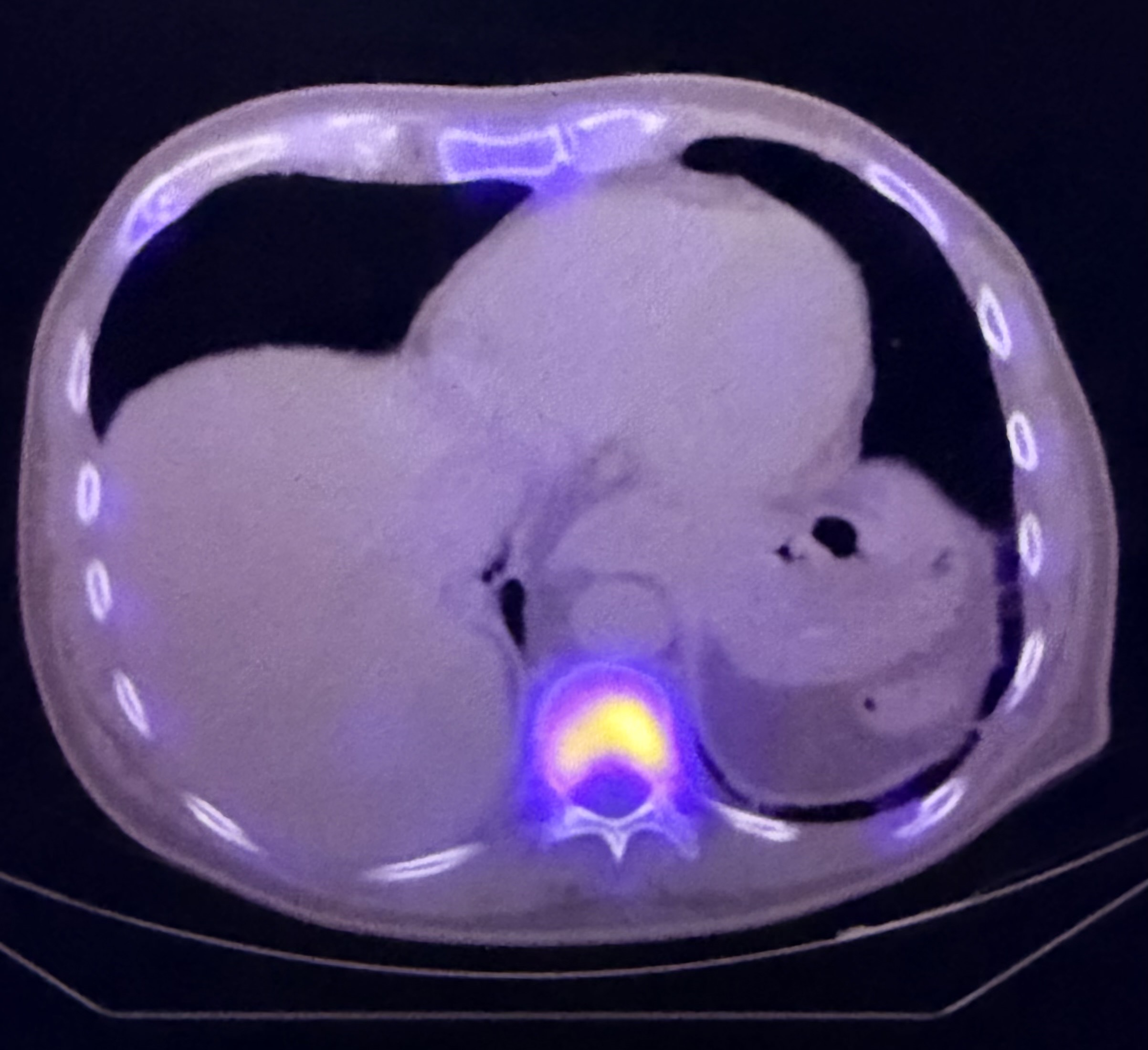
Detection usually needs a chain: suspicion, imaging or lab triage, tissue or cytology when needed, then pathology and molecular testing where it changes management.
Pros, cons and controversy
Benefits, harms and the false-positive problem.
The danger is delay from embarrassment or assuming pain is required. Most lumps are not cancer, but solid intratesticular masses need rapid workup.
Sources and trials